A peer-reviewed study published in the Journal of Alzheimer’s Disease examined whether bilingualism is associated with differences in brain structure among individuals with probable Alzheimer’s disease (AD). Using quantitative MRI volumetrics, researchers compared regional brain volumes in bilingual and monolingual patients matched for age and cognitive severity.
Background
Bilingualism has increasingly been recognized as a potential protective factor in individuals at risk for Alzheimer’s disease. Several studies suggest that bilingual individuals may experience a delay in the onset of clinical dementia symptoms despite comparable underlying neuropathology. One proposed explanation for this phenomenon is cognitive reserve—the brain’s ability to tolerate pathology while maintaining cognitive function.
Brain volume is a well-established structural marker of cognitive reserve. Larger regional brain volumes are generally associated with greater resilience to neurodegenerative disease.
Study Objective
The purpose of this proof-of-concept study was to compare MRI-measured brain volumes in bilingual versus monolingual individuals with probable Alzheimer’s disease who were matched for age and overall cognitive impairment.
Methods
Seventeen bilingual patients with probable Alzheimer’s disease were identified and matched to twenty-eight monolingual patients based on age at presentation and Mini-Mental State Examination (MMSE) scores. All bilingual participants were sequential bilinguals who learned English after age five and were functionally fluent in two languages prior to symptom onset.
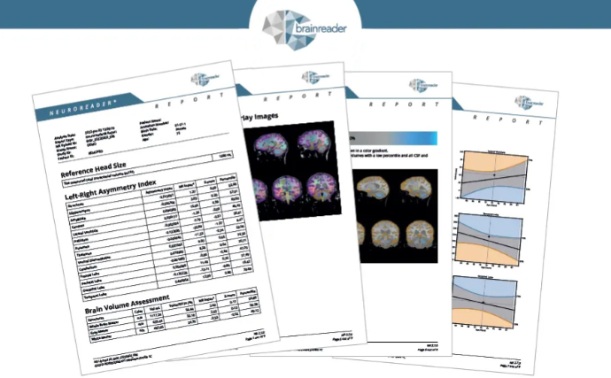
All participants underwent 3D volumetric T1 MRI scans on a 3.0 Tesla scanner. Brain volumes were quantified using Neuroreader®, an FDA-cleared automated brain volumetrics software. Forty-five brain structures were measured, including cortical, subcortical, ventricular, and brainstem regions.
Regional brain volumes were normalized to total intracranial volume and analyzed using statistical models adjusted for age, sex, and total intracranial volume.
Results
Bilingual individuals demonstrated larger brain volumes across numerous regions compared to monolingual individuals. Statistically significant differences were observed in the following structures:
- Brainstem
- Ventral diencephalon
Partial correlation analyses showed that bilingualism was significantly associated with larger volumes in:
- Brainstem
- Thalamus
- Ventral diencephalon
- Pallidum
Hippocampal volume showed a positive correlation with bilingualism, though this finding did not reach statistical significance. No brain regions were larger in the monolingual group, and no significant interaction was observed between bilingualism and MMSE scores on brain structure.
Interpretation
At comparable levels of cognitive impairment, bilingual individuals with Alzheimer’s disease exhibited greater preservation of specific subcortical and brainstem structures. These findings suggest that bilingualism may be associated with structural brain differences that reflect increased cognitive reserve.
Notably, several of the regions identified—such as the ventral diencephalon and brainstem—are not routinely emphasized in qualitative MRI interpretation, highlighting the value of quantitative volumetric analysis.
Discussion
Previous neuroimaging studies in cognitively normal bilingual individuals have demonstrated increased gray and white matter volumes in language-related and executive control networks. This study extends those findings into an Alzheimer’s disease population, suggesting that bilingualism may also be associated with preserved subcortical structures even after clinical dementia is established.
The ventral diencephalon and brainstem have both been implicated in Alzheimer’s disease pathophysiology and cognitive reserve. Preservation of these regions may contribute to delayed clinical expression of symptoms despite underlying neurodegeneration.
Limitations and Future Directions
This study was designed as a proof of concept and included a relatively small sample size. Future research is needed to evaluate longitudinal brain volume changes, incorporate functional and molecular imaging, and further explore regional specificity, including hippocampal subfields and language networks.
Conclusion
This study provides quantitative MRI evidence that bilingual individuals with Alzheimer’s disease demonstrate greater preservation of select brain structures compared to matched monolingual individuals. These findings support the hypothesis that bilingualism contributes to cognitive reserve and may play a role in delaying the clinical manifestation of Alzheimer’s disease.