Automated Brain Volumetric
Analysis From Routine MRI
Neuroreader® automatically analyzes MRI scans and
generates a quantitative volumetric brain report in
under 10 minutes.
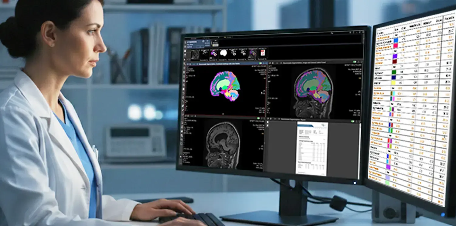
Automated Brain Volumetric Analysis
From Routine MRI
Neuroreader® automatically analyzes MRI scans and generates a quantitative volumetric brain report in under 10 minutes.
Neuroreader® is an FDA-cleared, CE-marked brain volumetric analysis software that transforms routine brain MRI into objective brain health insight. By precisely measuring brain structure and identifying patterns of atrophy, Neuroreader® helps clinicians better understand brain health over time, supporting earlier insight, improved differentiation, and confident clinical decision-making.
Developed by Brainreader, Neuroreader® is used globally by neuroradiologists, neurologists, and imaging centers as part of a brain health–first approach to neuroimaging.
Neuroreader® Capabilities

Comprehensive Brain Quantification
- Measures 83 brain volumes, including hippocampus, ventricles, lobes, and additional key structures
- Outputs volumes in mL, percentiles, and Z-scores
- Includes Total Intracranial Volume (mTIV) adjustment

Fast, Automated Results
- Entire brain processed in under 10 minutes
- No manual tracing required
- Automated segmentation enhances workflow efficiency

Normative Comparisons
Volumes are compared against an FDA-cleared normative database using:
- Age adjustment
- Sex adjustment
- Head size (mTIV) normalization

Longitudinal Tracking
- Trend charts across repeat MRI studies
- Whole-brain and regional volume change visualization
- Objective monitoring of disease progression

Asymmetry analysis for TBI
- Left-Right Asymmetry Index (13 structures)
- Supports evaluation of traumatic brain injury and focal structural changes
- Enhances reproducibility in subtle cases

Flexible Deployment
- PACS integration
- Secure web portal access
- Designed for clinical and enterprise environments
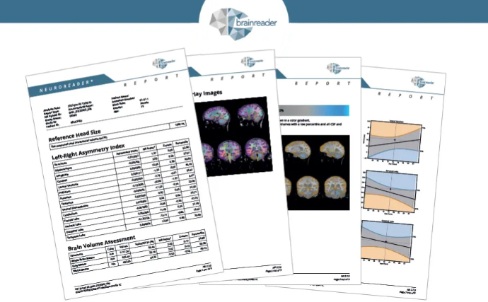
Inside the Neuroreader® Report
The Neuroreader® report combines quantitative measurements with visual segmentation to assist clinicians in evaluating brain structure volumes.
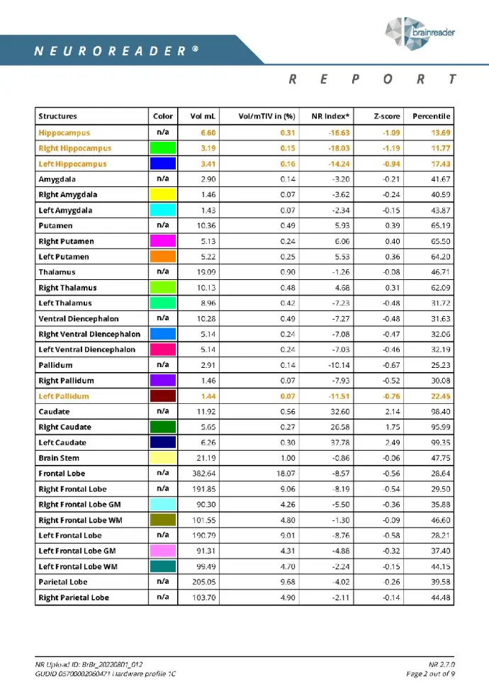
Brain Volume Assessment Table
Quantified Data on 83 Brain Structures
- Key Metrics:
- Structure name
- Measured volume (mL)
- Z-score (standard deviations from the mean)
- Percentile rank (relative to normal population)
- NR Index (aggregated health score)
- All brain matter volumes with a percentile lower than 25% and all CSF structures with a percentile over 75% are shown in orange
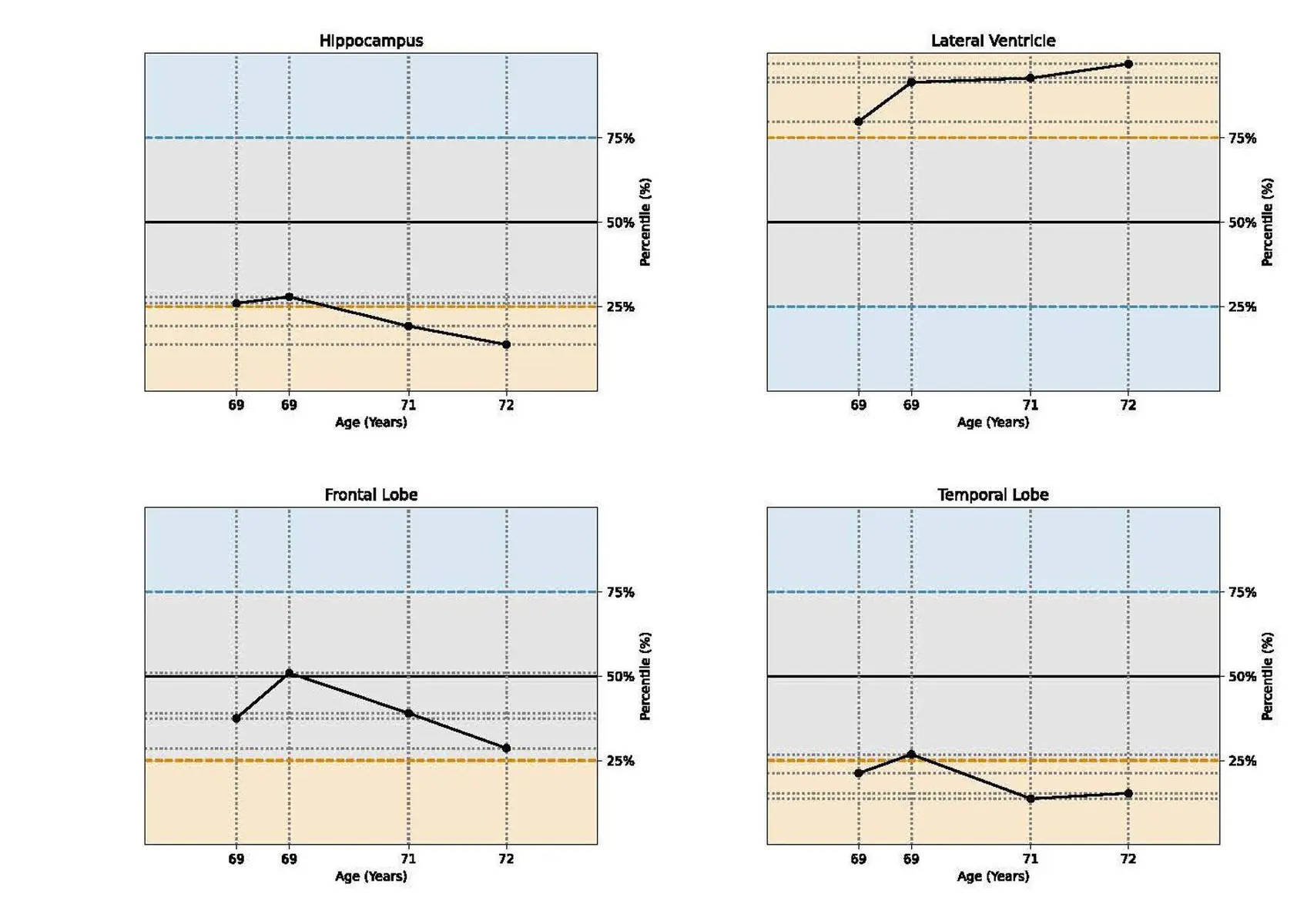
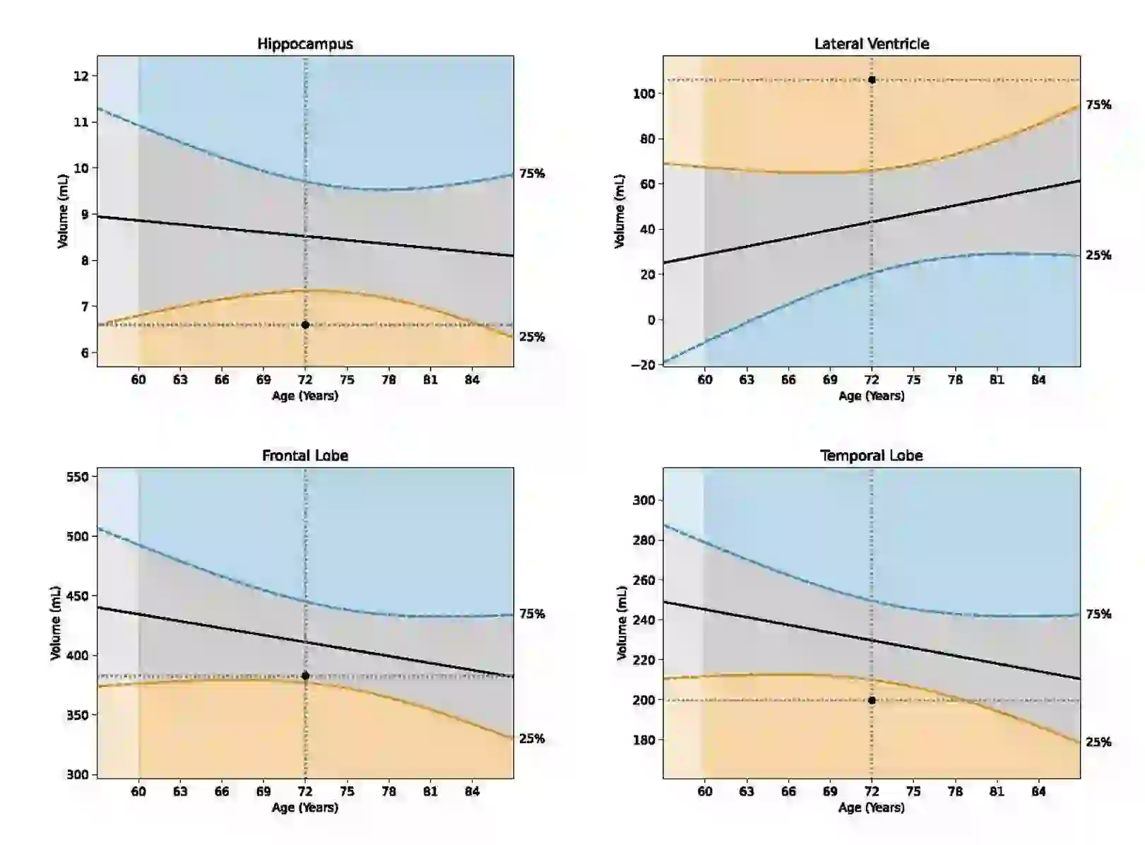
Longitudinal Analysis
Tracking Structural Change Over Time
- Trend graphs showing change in key structures (e.g., hippocampus, ventricles)
- Timepoints from multiple scans (e. g., every 6–12 months)
- Percent change vs. baseline
- Enables clinicians to objectively monitor disease progression.
- Critical for evaluating therapy response or cognitive decline trends.
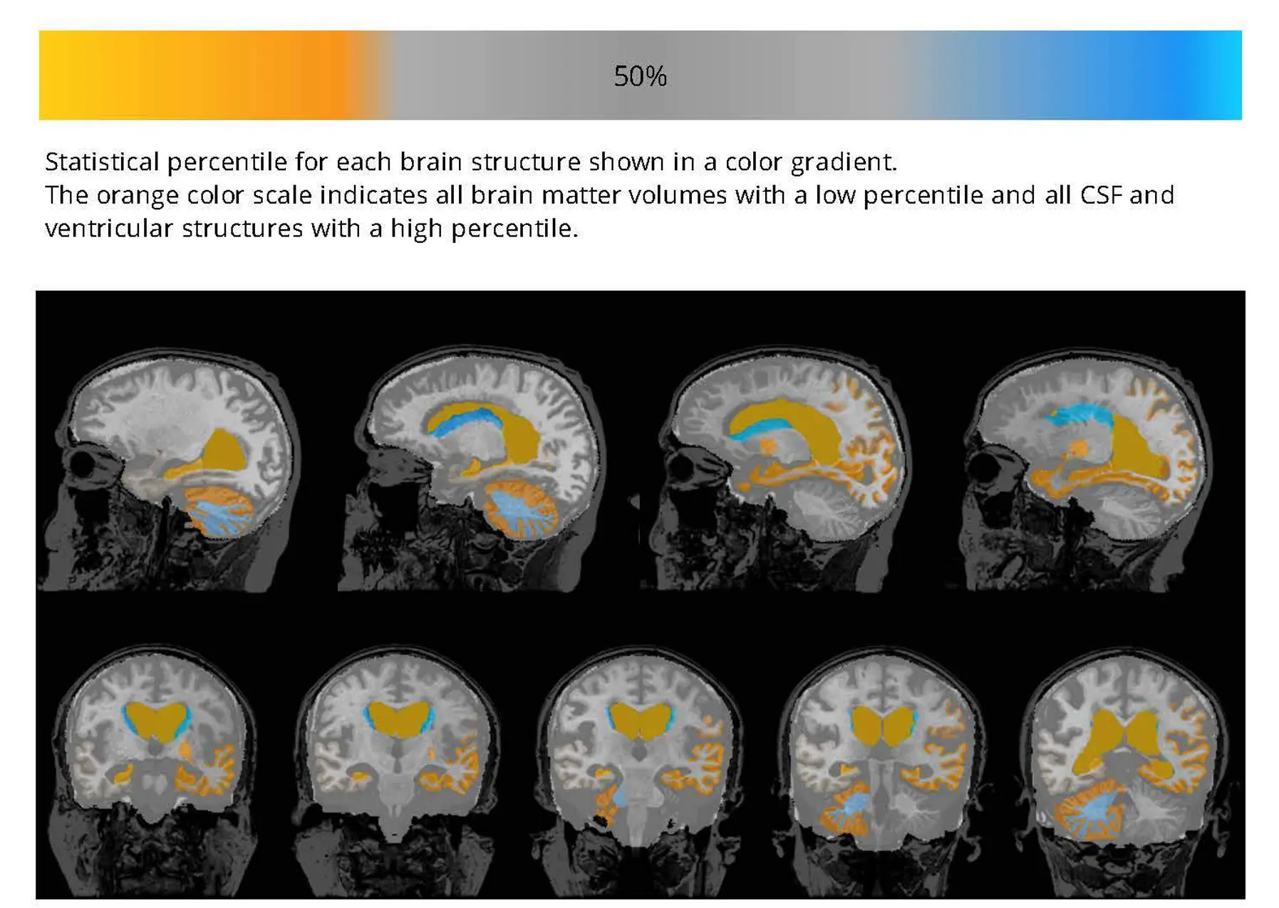
Brain Heat Maps
Visualizing Areas of Concern
- Color-coded overlays:
- Blue = above normal
- Orange = below normal (atrophy)
- Interactive percentile visualization of brain structures
- Offers an intuitive, patient-friendly way to visualize neurodegeneration
- Ideal for neurologist-patient discussions and for progress visualization in follow-up scans.
Charts
Visualizing Atrophy Trends
- Bar charts summarizing Z-scores or percentile values
- Comparative visualization of key brain regions
- Provides at-a-glance understanding of which regions are abnormal
- Ideal for summarizing case data in reports or research studies
Clinical Applications of Automated Brain Volumetrics
Dementia & Neurodegeneration
Volumetric MRI supports evaluation of patients with suspected cognitive impairment.
Neuroreader® provides:
- Objective hippocampal measurements
- Whole-brain atrophy assessment
- Regional structural comparisons
Demographic-adjusted analyses assist clinicians in identifying early biomarkers of neurodegeneration.
Traumatic Brain Injury (TBI)
Subtle structural changes may not be visually apparent on routine MRI.
Neuroreader® provides:
- Objective volumetric measurements
- Left-right asymmetry indices
- Standardized segmentation
This supports interpretation in TBI and mesial temporal sclerosis cases.
Longitudinal Monitoring
Across repeat studies, Neuroreader® enables visualization of:
- Percentile shifts
- Whole-brain volume change
- Regional structural progression
Longitudinal volumetrics may assist clinicians in monitoring disease progression and evaluating treatment response.
How Neuroreader® Fits Into the MRI Workflow
Neuroreader® is designed to integrate into routine brain MRI protocols with minimal disruption.
MRI Acquisition
A standard 3D T1 sequence is acquired on a 1.5T or 3.0T scanner without contrast.
Secure Upload or PACS Routing
Images are uploaded securely to the Neuroreader® system or routed via PACS integration.
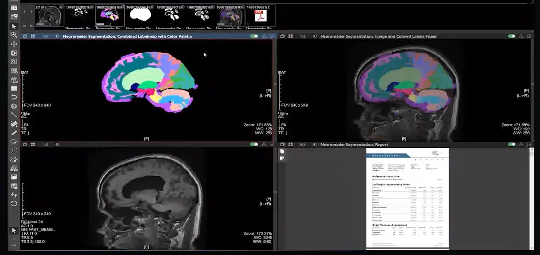
Automated Segmentation
The software automatically segments and quantifies 83 brain structures.
Report Generation
A customized volumetric report is generated and returned in under 10 minutes.
Manual Segmentation vs. Neuroreader®
Manual Segmentation
- Time-intensive
- Inter-reader variability
- Limited structures
- Subjective interpretation
- Labor intensive
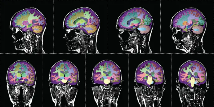
Neuroreader®
- <10 minute automated report
- Standardized segmentation
- 83 brain volumes
- Quantitative percentiles & Z-scores
- Fully automated
Neuroreader® brings efficiency and objectivity to volumetric MRI analysis.
Seemless Integration
No Downtime. No Hardware. No Disruption.
- PACS Integration: Works directly within your Infinitt, or other PACS viewer, with one-click access to Neuroreader® reports.
- Secure Web Portal: Upload studies directly via browser for remote or multi-site use
- Automated Processing: Reports automatically returned to your PACS, without interrupting routine MRI workflows
- Clinician-Friendly: Radiologists validate images and order Neuroreader® with a single mouse click
- Fully Compliant: HIPAA, FDA, and CE certified
- Compatible with all major MRI scanner brands (Siemens, GE, Philips)
- Uses 3D T1 sequence (1–1.2 mm slice thickness)
- Configured and running in under 5 minutes
Regulatory Compliance
& Clinical Certification
Designed to meet U.S. and international medical device standards.
All PHI data is handled securely in accordance with healthcare regulatory requirements.

Request a Sample Neuroreader® Report
See how automated brain volumetrics can enhance diagnostic confidence and streamline MRI interpretation.