Radiologists and neurologists have long relied on MRI as the gold standard for evaluating brain structure. Yet, conventional interpretation often depends heavily on the reader’s expertise and visual judgment. Subtle changes can be overlooked, and results may vary between clinicians.
This is where automated volumetric MRI like Neuroreader is changing the field by shifting brain imaging from primarily subjective interpretation to objective, quantifiable analysis.
The Challenge of Subjective Interpretation
MRI scans provide exceptional detail, but manual interpretation is limited by:
- Variability between readers: Two radiologists may describe the same scan differently.
- Time-intensive processes: Manual segmentation of brain structures can take hours.
- Difficulty detecting subtle changes: Early neurodegeneration may be too subtle for visual detection.
For imaging directors managing workflow efficiency, these challenges can slow reporting, increase costs, and reduce consistency across teams.
How Automated Volumetric MRI Works
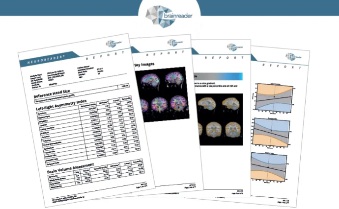
Volumetric brain analysis software applies advanced algorithms to quantify the volume of individual brain structures. Instead of relying only on visual impressions, clinicians receive numerical values and comparisons to normative datasets.
With MRI segmentation automation, more than 60 structures — including the hippocampus, ventricles, and cortical lobes — can be measured in under 10 minutes. This turns what was once a subjective process into a reproducible, objective report.
Why Objectivity Matters in Diagnosis
Shifting from subjective reads to MRI objectivity in diagnosis brings several clinical advantages:
- Earlier detection: Quantitative reports can identify atrophy years before symptoms.
- Consistency across readers: Automated methods reduce variability and subjectivity.
- Tracking progression: Longitudinal reports make it possible to measure change over time.
- Improved confidence: Clinicians can rely on reproducible data to support treatment planning.
Seamless Integration into Workflow
Modern volumetric brain analysis software is designed to work within existing PACS or through secure web access. Reports are generated quickly, without adding scan time or requiring new hardware. This makes automated volumetric MRI a practical addition to routine imaging protocols.
The Future of Brain Imaging
As radiology departments face growing demand and pressure to improve efficiency, tools that deliver objective, automated volumetric MRI data will play a greater role. For neurologists and imaging directors, this means the opportunity to provide earlier interventions, standardize reporting, and strengthen clinical decision-making.