Why Accurate Detection of Mesial Temporal Sclerosis Matters
Mesial temporal sclerosis (MTS) is one of the most common and surgically treatable causes of temporal lobe epilepsy (TLE). Accurate identification of MTS is critical because:
- Patients with confirmed MTS are more likely to achieve seizure freedom after temporal lobectomy
- MRI-negative or subtle cases often require invasive intracranial EEG monitoring
- Missed or delayed diagnosis can prevent patients from accessing potentially curative surgery
However, visual MRI interpretation alone is limited, particularly when hippocampal atrophy is subtle or confounded by age-related brain volume loss, prior injury, or diffuse atrophy.
Study Overview: Automated Volumetric MRI vs Radiologist Interpretation
A peer-reviewed study published in the American Journal of Neuroradiology evaluated whether automated hippocampal volumetry using FDA-cleared Neuroreader® software could more accurately identify MTS compared to standard neuroradiologist interpretation.
Study Design
- 71 adult patients with medically refractory temporal lobe epilepsy
- All underwent preoperative MRI followed by temporal lobectomy
- Pathology-confirmed diagnosis served as the reference standard
- MRI scans were analyzed using Neuroreader® automated segmentation
- Results were compared to routine clinical neuroradiology reports
Hippocampal volume was measured using:
- Percentage of hippocampal volume relative to total intracranial volume (%Vol)
- Neuroreader Index (NRI) — an age- and sex-adjusted normative comparison
Key Results: Neuroreader® Significantly Improves Sensitivity
Diagnostic Performance
| Method | Sensitivity | Specificity |
| Radiologist MRI Interpretation | 50% | 87% |
| Automated Hippocampal %Vol | 89% | 71% |
| Neuroreader Index (NRI) | 89% | 78% |
Automated volumetric analysis identified mesial temporal sclerosis significantly more accurately than radiologist interpretation alone (P < 0.0001).
Optimal Diagnostic Thresholds
- Hippocampal %Vol: ≤ 0.19%
- Neuroreader Index: ≤ 3.8
Volumes below these thresholds were strongly suggestive of mesial temporal sclerosis.
Why Automated Volumetry Detects What the Human Eye Misses
Radiologic diagnosis of MTS traditionally relies on:
- Visual hippocampal volume loss
- T2 hyperintensity
However, pathology-proven MTS frequently exists without obvious signal abnormality, especially in early or subtle disease.
Automated volumetry provides:
- Objective measurement rather than subjective assessment
- Normalization for age, sex, and head size
- Consistency across readers and institutions
- Improved detection in cases labeled “normal” or “equivocal” on MRI
This makes volumetric MRI particularly valuable when:
- MRI findings are subtle
- EEG findings are lateralizing but imaging is inconclusive
- Avoiding invasive diagnostic procedures is a priority
Clinical Impact for Epilepsy Care Teams
For Neuroradiologists
- Increased confidence in identifying subtle hippocampal atrophy
- Reduced false-negative MRI reports
- Objective data to support or refute suspected MTS
For Neurologists & Epileptologists
- Earlier identification of surgical candidates
- More informed decisions about invasive EEG monitoring
- Better patient counseling and care planning
For Patients
- Faster diagnosis
- Fewer unnecessary invasive procedures
- Improved access to potentially curative epilepsy surgery
Why Neuroreader® Is Clinically Relevant
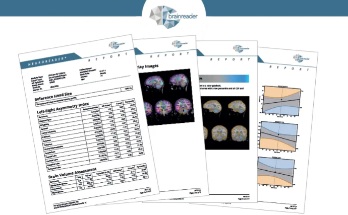
Neuroreader® is an FDA-cleared, CE-marked automated brain volumetrics software that integrates directly into clinical MRI workflows and produces results in under 10 minutes.
Key advantages include:
- Automated segmentation of hippocampi and other brain regions
- Age- and sex-adjusted normative comparison
- Proven performance across neurodegenerative disease, TBI, and epilepsy
- Designed for routine clinical use, not research-only workflows
Neuroreader® has been validated across multiple peer-reviewed studies and is used globally to support objective brain MRI interpretation
Brainreader Information
Conclusion: A New Standard for Detecting Mesial Temporal Sclerosis
This study demonstrates that automated hippocampal volumetry using Neuroreader® outperforms subjective MRI interpretation for detecting mesial temporal sclerosis in temporal lobe epilepsy.
With validated thresholds and pathology-confirmed accuracy, volumetric MRI:
- Improves diagnostic sensitivity
- Reduces variability
- Enhances clinical decision-making
- Supports earlier, more confident epilepsy surgery evaluation
Automated brain volumetrics are no longer optional — they are becoming essential in modern epilepsy imaging.