A 2019 study in the Journal of Alzheimer’s Disease found that people with a history of traumatic brain injury (TBI) who present with cognitive complaints show measurable, region-specific brain atrophy on volumetric MRI—most notably in the ventral diencephalon, putamen, pallidum, and thalamus—while the hippocampus is relatively spared. That pattern differs from typical Alzheimer’s disease, and several affected regions correlate with lower MMSE/MoCA scores. Clinically, this supports using automated MRI volumetrics to add objective evidence when evaluating post-TBI cognitive decline and to help differentiate probable etiologies.
Key takeaways from the study
- Who was studied: 40 patients (mean age ~68) with documented TBI and cognitive symptoms underwent 3D T1 MRI with FDA-cleared volumetric analysis.
- What was found: Greatest volume loss was seen in ventral diencephalon, putamen, pallidum, with smaller effects in temporal lobes and brainstem; hippocampus showed the least atrophy despite correlating with cognition.
- Why it matters: Lobar atrophy correlated with MMSE/MoCA, supporting volumetrics as a sensitive adjunct for assessing cognition after TBI. The relative hippocampal sparing suggests a pattern less consistent with classic Alzheimer’s, aiding differential consideration.
How this translates to practice
Conventional reads can miss subtle, spatially distributed atrophy. Automated volumetric MRI provides:
-
Objective, reproducible measurements adjusted for age, sex, and head size (normative comparisons), helping clinicians quantify “how much” and “where” atrophy is occurring.
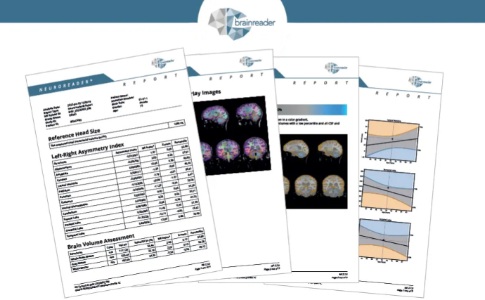
Brainreader Information -
Fast turnaround—a volumetric report can be generated in under 10 minutes, fitting real clinical workflows and reducing reporting subjectivity.
Brainreader Information
Brainreader Information -
Regulatory assurance (FDA-cleared, CE-marked) and seamless access via web, PACS, or mediation server—so teams can adopt without heavy IT lifts.
Brainreader Information
Brainreader Information
Bonus for TBI workflows: Brainreader’s configurable reporting includes an Asymmetry Report, often useful when TBI or mesial temporal sclerosis is suspected.
Brainreader Information
Bottom line
For patients with post-TBI cognitive impairment, MRI volumetrics can uncover a non-AD atrophy signature and provide quantitative evidence that complements neuropsychological testing—supporting clearer clinical conversations and more confident care planning.
Interested in adding objective volumetrics to your TBI workups or neurology/radiology service line? We can help you plug automated MRI analysis into your existing workflow in minutes and return a structured report right to PACS.