Neurology study links exercise, metabolic health, and preserved brain volume
A research study published on April 13, 2022, in Neurology—the medical journal of the American Academy of Neurology—reports that physical activity improves cognitive brain function by reducing body mass index (BMI) and improving insulin metabolism. These metabolic improvements were shown to mediate the relationship between physical activity and preserved gray matter volume, a key marker of brain health.
How Does Exercise Protect Brain Health?
Previous research has consistently shown that larger gray matter volume is associated with better cognitive function and may help protect against dementia. Gray matter plays a critical role in information processing, memory, and decision-making.
This new study expands on earlier findings by demonstrating that insulin resistance and BMI influence the relationship between physical activity and brain gray matter volume. In other words, exercise may help preserve brain structure in part by improving metabolic health.
Study Design and Participants
The study included 134 cognitively healthy adults with an average age of 69 years. Key components of the research included:
- A self-reported physical activity survey covering the previous 12 months
- Brain imaging scans to measure gray matter volume
- Assessments of glucose metabolism, insulin resistance, and BMI
By combining metabolic data with MRI-based brain measurements, researchers were able to evaluate how physical activity influences brain structure through physiological pathways.
Key Takeaways from the Study
“Exercise has often been called food for the brain, with many studies showing the benefit of exercise for improving brain health and reducing the risk of dementia.”
According to Dr. Zablow, former assistant professor at the University of California San Diego Medical School:
“This current research study states that physical activity improves cognitive brain function by reducing BMI and improving insulin metabolism. Improvement in weight control can limit the rate of brain volume loss, a known risk factor for dementia.”
He further noted:
“This study will help physicians reinforce the importance of regular exercise in reducing BMI as a low-cost means of limiting cognitive decline.”
Why Glucose Metabolism Matters for the Brain
Glucose metabolism is essential for brain function. The brain relies on glucose to generate adenosine 5′-triphosphate (ATP)—a molecule critical for:
- Maintaining neuron health
- Supporting synaptic activity
- Producing neurotransmitters
Reduced glucose metabolism in the brain is commonly observed in people with dementia, underscoring the importance of metabolic health in preserving cognition.
Brain Volume, Aging, and Cognitive Decline
Gray matter development peaks between ages 2–3 years, after which total volume gradually declines in certain brain regions. While density may increase during development, brain tissue loss later in life is strongly associated with cognitive decline.
Although total brain volume has only a weak correlation with intelligence, progressive brain atrophy is a significant contributor to reduced cognitive ability with aging.
MRI-based volumetric analysis allows clinicians to quantify these changes objectively and monitor brain health over time.
Expert Perspective: Why These Findings Matter
In commentary provided to Medical News Today, Dr. Raeanne Moore, associate adjunct professor of psychiatry at UC San Diego, stated:
“The literature clearly demonstrates that cardiovascular risk factors are associated with cognitive decline and risk for Alzheimer’s disease and related dementias.”
She added:
“This study adds to the growing body of research on the positive benefits of staying active on brain health, especially as we age.”
Dr. Moore concluded:
“Studies investigating subtle brain changes prior to the development of dementia are critical to optimizing brain health and staving off cognitive decline.”
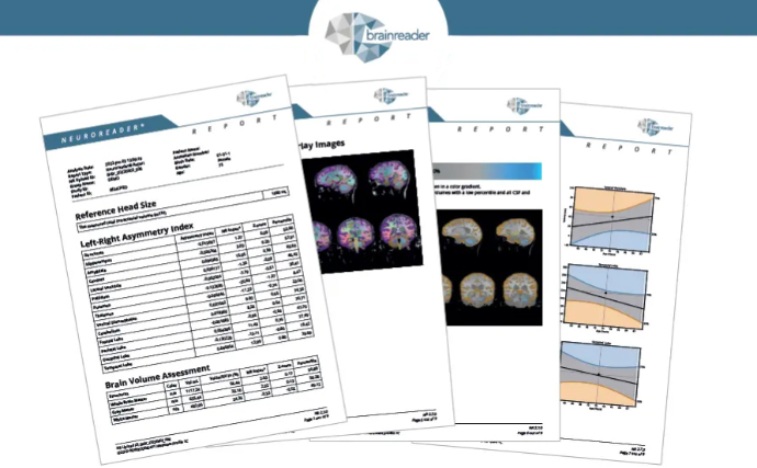
Measuring Brain Volume with Quantitative Neuroimaging
Advances in quantitative neuroimaging now allow clinicians to measure total brain and regional volumes from routine MRI scans. Tools like Neuroreader® provide objective, reproducible brain volume data to support research, clinical assessment, and longitudinal monitoring.