Event Date: June 15, 2017
Published: May 25, 2017
Source: Silicon Valley Health Institute
Overview: Brain Health and Imaging
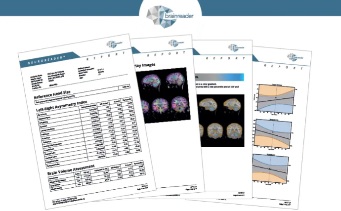
Quantitative neuroimaging is transforming how clinicians and researchers understand brain health. In this expert presentation, Cyrus A. Raji, MD, PhD explores how MRI-based brain volumetrics provide objective insights into neurological and neuropsychiatric disorders—and how brain volume may function as a measurable vital sign for cognitive health.
Brain volume measurements derived from MRI scans reveal structural changes associated with conditions such as Alzheimer’s disease, depression, and traumatic brain injury (TBI). When brain volumes fall outside normal ranges, they often correlate with impaired neuronal function and increased risk of cognitive decline.
Drawing on more than a decade of peer-reviewed research, Dr. Raji demonstrates that brain volumes are not static. Instead, they can change in response to modifiable lifestyle factors, including physical activity, obesity, and dietary habits. These findings have significant implications for early detection, prevention strategies, and long-term brain health preservation.
This presentation outlines:
- How quantitative MRI volumetrics measure regional brain structure volumes
- Why brain volume can be considered a biological marker of brain health
- Evidence linking lifestyle factors to structural brain changes
- A forward-looking framework for integrating volumetric imaging into preventive medicine
Why Quantitative Neuroimaging Matters
Traditional MRI interpretation relies heavily on visual assessment, which may miss subtle or diffuse patterns of brain atrophy. Quantitative neuroimaging enhances clinical insight by providing:
- Objective, reproducible measurements of brain structures
- Early detection of volume changes associated with neurodegenerative and neuropsychiatric disorders
- Data-driven support for prevention and monitoring strategies
- A foundation for precision medicine approaches in neurology and psychiatry
This shift from subjective interpretation to data-driven brain health imaging represents a major advancement in modern medicine.
About Dr. Cyrus A. Raji, MD, PhD

Dr. Cyrus A. Raji is a physician-scientist specializing in neuroradiology and neuroimaging research. At the time of this presentation, he was a neuroradiology clinical fellow and a member of the NIH-funded T32 research program at the UCSF Department of Radiology and Biomedical Imaging.
Education and Training
- MD/PhD, Medical Scientist Training Program, University of Pittsburgh School of Medicine (2010)
- Postdoctoral research, Department of Psychiatry, University of Pittsburgh
- Transitional medicine internship, UPMC Mercy Hospital (2011–2012)
- Diagnostic radiology residency, UCLA Medical Center
Research and Funding
Dr. Raji has received research funding from:
- American Heart Association
- Radiological Society of North America (RSNA)
- Foundation of the American Society of Neuroradiology
He has authored 30+ peer-reviewed publications in leading journals including Neurology, Human Brain Mapping, American Journal of Preventive Medicine, and American Journal of Neuroradiology. His work focuses on applying structural and functional neuroimaging to neuropsychiatric disorders, head trauma, and cognitive decline.
Featured Short Presentation: 3D Printing in Healthcare
3D Printing in Healthcare
Presented by: Hui Jenny Chen, MD
Although 3D printing technology has existed for decades, recent advances in affordability and materials have accelerated its adoption across healthcare. This short presentation explores how medical 3D printing is being used in applications such as orthodontics, hearing aids, and personalized medical devices—delivering direct benefits to patients.
About Jenny Chen, MD

Dr. Jenny Chen is a neuroradiologist and the Founder and CEO of 3DHEALS, a platform dedicated to building and educating the healthcare 3D printing ecosystem. Her professional interests include:
- Medical education
- 3D printing in healthcare
- Artificial intelligence in medical imaging
She also serves as an adjunct clinical faculty member in the Department of Radiology at Stanford Health Care.
Key Takeaway
Quantitative brain imaging represents a paradigm shift in how clinicians assess, monitor, and protect brain health. By combining MRI volumetrics with lifestyle and clinical data, researchers like Dr. Cyrus Raji are redefining prevention, early detection, and personalized care for neurological disorders.