Chronic traumatic encephalopathy (CTE) is a neurodegenerative disorder associated with repetitive head trauma, commonly affecting former football players and others with a history of concussions. Until now, CTE could only be definitively diagnosed after death through brain autopsy.
A UCLA-led case study suggests that MRI-based brain volumetric analysis may offer a potential path toward identifying CTE-related brain changes in living patients.
UCLA Researchers Identify CTE-Like Brain Changes Using MRI
Using advanced MRI analysis software, UCLA physicians identified abnormal shrinkage in key brain regions in a former football player experiencing cognitive and behavioral symptoms.
Importantly, the pattern of brain atrophy observed on MRI closely matched findings previously seen in autopsy-confirmed CTE cases, raising the possibility that MRI could support earlier, non-invasive assessment of CTE.
While this report is based on a single patient and remains preliminary, it represents an important step toward improving how clinicians evaluate long-term brain health after repetitive head injury.
Why Early CTE Detection Matters
Currently, CTE can only be diagnosed through post-mortem brain examination, limiting both clinical care and research.
“Having an MRI-based technique for detecting this pattern of brain changes would help us a lot in assessing the brain health of athletes and others with histories of concussions,”
— Dr. David Merrill, UCLA Semel Institute for Neuroscience and Human Behavior
An MRI-based approach could:
- Enable earlier clinical evaluation
- Improve monitoring of patients with concussion history
- Accelerate CTE research and therapeutic development
Case Background: Former Football Player With Cognitive Symptoms
The patient, referred to by the pseudonym “Bob Smith,” was a former high-school football fullback who played extensively on offense, defense, and special teams. Over three seasons, he experienced hundreds of head impacts, including at least one concussion involving loss of consciousness.
In his late 30s, Smith developed:
- Mood instability
- Attention and impulse-control difficulties
- Executive function impairment
Standard neuropsychological testing confirmed these deficits, though memory remained intact. Conventional MRI ruled out Alzheimer’s disease, stroke, and dementia.
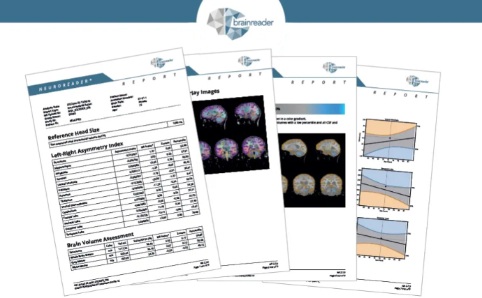
MRI Volumetric Analysis Reveals Progressive Brain Atrophy
While routine MRI showed no obvious neurodegenerative disease, advanced MRI volumetric analysis identified:
- Abnormally low volumes in the brainstem
- Reduced volume in the ventral diencephalon
- Frontal lobe atrophy
- Progressive worsening over a four-year interval
Overall, the patient lost approximately 14% of total gray-matter volume during that time.
“The specific areas that were abnormally low in their volumes helped us understand that this patient’s cognitive symptoms were likely due to his history of traumatic brain injury,”
— Dr. Cyrus Raji, Ronald Reagan UCLA Medical Center
Advantages of MRI-Based CTE Assessment
Researchers note that other investigational methods for diagnosing CTE in living patients—such as tau PET imaging—require radioactive tracers and are costly.
An MRI-based approach offers key advantages:
- Non-invasive
- No radiation exposure
- Lower cost
- Widely available in clinical practice
According to Dr. Merrill, this makes MRI a safer and more accessible tool for both patients and researchers.
Next Steps in CTE Research
The research team plans to conduct larger studies involving individuals with histories of repetitive head trauma to determine whether MRI volumetric analysis can reliably detect CTE-associated brain changes across broader populations.
The study was authored by Drs. Cyrus Raji and David Merrill and published online August 23 in the American Journal of Geriatric Psychiatry. Additional contributors included researchers from UCLA and UC Davis.
Key Takeaways for Clinicians and Researchers
- MRI volumetric analysis may reveal CTE-like brain atrophy patterns in living patients
- Early findings align with known post-mortem CTE pathology
- Larger studies are needed before clinical adoption
- MRI could become a safer, scalable tool for CTE assessment and research