Blog
Ekspertindsigt i AI-drevet hjernebilleddannelse og klinisk innovation
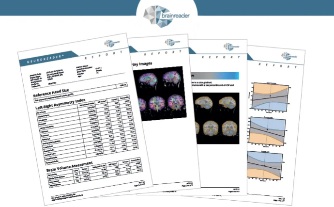
Request a Sample Neuroreader® Report
See how automated brain volumetrics can enhance diagnostic confidence and streamline MRI interpretation.