Authors:
Cyrus A. Raji, MD, PhD; David A. Merrill, MD, PhD; Jorge R. Barrio, PhD; Bennet Omalu, MD, MPH; Gary W. Small, MD
Journal: American Journal of Geriatric Psychiatry (October 2016)
DOI: 10.1016/j.jagp.2016.07.018
Overview
This peer-reviewed case study describes progressive focal gray matter volume loss detected with longitudinal volumetric MRI in a 51-year-old former high school football player with a history of repetitive head trauma and concussions. The findings suggest a potential MRI volumetric signature of chronic traumatic encephalopathy (CTE) and highlight the clinical value of automated brain volumetrics in suspected traumatic brain injury (TBI).
Case Summary: Former High School Football Player With Cognitive Decline
The patient presented with six years of progressive cognitive and mood decline, including memory impairment, attention deficits, executive dysfunction, and depressive symptoms. His history included:
- Multiple repetitive head impacts during high school football
- At least one concussion with loss of consciousness
- Progressive occupational impairment
- Prior diagnoses of ADHD and bipolar II disorder
Neuropsychological testing showed executive dysfunction with preserved memory, suggesting a non-Alzheimer’s pattern of cognitive decline.
Neuroimaging Methods: Longitudinal Volumetric MRI
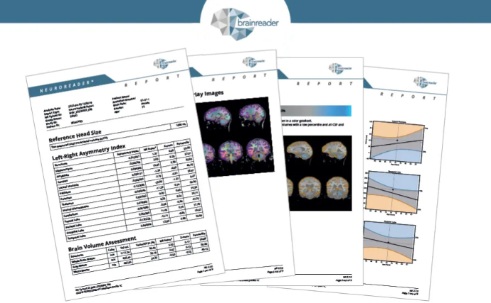
Two 3T MRI scans (2012 and 2016) were analyzed using FDA-cleared automated volumetric MRI software (Neuroreader®). Imaging included:
- 3D T1-weighted volumetric sequences
- T2, FLAIR, and gradient echo sequences
- Longitudinal volumetric comparison over four years
Volumetric MRI enabled objective quantification of regional brain atrophy, surpassing the sensitivity of visual inspection alone.
Key MRI Findings
Progressive Regional Brain Atrophy
Longitudinal volumetric analysis revealed:
- 14% total gray matter volume loss
-
Marked atrophy in:
- Brainstem
- Ventral diencephalon
- Frontal lobes
- Preserved hippocampal and temporal lobe volumes, arguing against Alzheimer’s disease and frontotemporal dementia
Supporting Structural MRI Findings
Additional MRI features consistent with prior traumatic brain injury included:
- Old petechial hemorrhages in frontal and temporoparietal white matter
- Areas of gliosis
- Cavum septum pellucidum, a finding previously associated with contact-sport–related CTE
Correlation With Molecular Imaging
The regional distribution of volumetric atrophy closely matched tau and amyloid PET signal patterns (FDDNP-PET) observed in separate suspected CTE cases, aligning with known tau deposition patterns in autopsy-confirmed CTE.
Clinical Significance
This case represents one of the first documented examples of longitudinal regional brain atrophy in a former high school football player with suspected CTE. While definitive diagnosis of CTE remains post-mortem, the combination of:
- Progressive executive dysfunction
- Frontal and brainstem atrophy
- Preserved hippocampal volume
- History of repetitive head trauma
supports the likelihood of CTE over alternative neurodegenerative conditions.
Implications for Volumetric MRI in CTE and TBI
If replicated in larger cohorts, these findings suggest that volumetric MRI may provide a reproducible, non-invasive biomarker for suspected CTE, with the added benefit of:
- Ruling out Alzheimer’s disease and frontotemporal dementia
- Supporting longitudinal disease monitoring
- Enhancing diagnostic confidence in patients with complex neuropsychiatric presentations
Automated volumetric MRI tools such as Neuroreader® allow rapid, standardized brain structure quantification and are increasingly relevant in both clinical neurology and neuroradiology workflows.
Conclusion
This case highlights the potential role of quantitative MRI volumetrics in identifying characteristic patterns of brain atrophy associated with suspected chronic traumatic encephalopathy. As contact sports participation continues, baseline and longitudinal volumetric MRI, combined with neuropsychological assessment, may become essential tools for early detection, monitoring, and clinical decision-making in traumatic brain injury.