
Similar Symptoms. Different Diseases.
Alzheimer's disease (AD) and frontotemporal dementia (FTD) are two of the most common causes of dementia, yet they affect the brain in different ways and often present with overlapping symptoms.
A patient experiencing memory loss, personality changes, difficulty finding words, or changes in executive function may initially appear to fit more than one diagnosis. While clinical history, neurological examination, cognitive testing, and laboratory evaluation remain the foundation of assessment, imaging also plays an important role in understanding structural changes within the brain.
Because Alzheimer's disease and frontotemporal dementia typically affect different brain regions, quantitative brain MRI can provide objective volumetric measurements that complement conventional MRI interpretation and support the overall clinical evaluation.
Understanding Alzheimer's Disease
Alzheimer's disease is the most common cause of dementia worldwide.
It is characterized by progressive degeneration of brain tissue, particularly within structures involved in memory formation and learning.
Common symptoms include:
- Progressive memory loss
- Difficulty learning new information
- Problems recalling recent conversations
- Confusion regarding time or place
- Difficulty completing familiar tasks
- Gradual decline in cognitive function
Although Alzheimer's disease primarily affects memory during its early stages, additional cognitive functions often become involved as the disease progresses.
Understanding Frontotemporal Dementia (FTD)
Frontotemporal dementia represents a group of neurodegenerative disorders that primarily affect the frontal and temporal lobes of the brain.
Unlike Alzheimer's disease, FTD frequently begins with changes in behavior, personality, judgment, or language rather than memory.
Patients may experience:
- Personality changes
- Reduced empathy
- Poor judgment
- Behavioral disinhibition
- Difficulty speaking or understanding language
- Executive dysfunction
Because these symptoms can resemble psychiatric disorders or overlap with Alzheimer's disease, accurate clinical evaluation is essential.
Why Differentiation Can Be Challenging
Although Alzheimer's disease and frontotemporal dementia affect different brain regions, patients may present with similar symptoms—particularly during the early stages of disease.
For example:
- Both conditions may involve memory complaints.
- Both may present with cognitive decline.
- Both can affect language.
- Both can interfere with daily functioning.

For clinicians, the challenge is not simply recognizing cognitive impairment, but understanding which neurodegenerative process may be contributing to the patient's symptoms.
A comprehensive evaluation typically includes:
- Medical history
- Neurological examination
- Cognitive assessment
- Laboratory testing
- Brain MRI
- Additional imaging when clinically appropriate
Understanding Mild Cognitive Impairment (MCI): Why Early Imaging Matters
The Role of Brain MRI
Conventional MRI remains one of the most important imaging tools in evaluating patients with cognitive decline.
MRI allows clinicians to assess:

Brain anatomy

Tissue integrity

Patterns of atrophy

Alternative causes of symptoms
However, subtle differences in regional brain volume may be difficult to objectively quantify through visual assessment alone.
This is where quantitative brain volumetry can provide additional insight.
What Is Quantitative MRI (qMRI) and Why Does It Matter?
How Quantitative Brain MRI Adds Objective Information
Quantitative MRI analyzes routine high-resolution 3D T1-weighted MRI studies to objectively measure brain structures.
Neuroreader® automatically segments and quantifies 83 brain regions in less than 10 minutes, providing standardized volumetric measurements that complement physician interpretation.
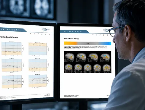
These measurements include:
- Regional brain volumes
- Whole-brain volume
- Left-right asymmetry indices
- Age-adjusted percentile comparisons
- Longitudinal volume tracking
Rather than replacing clinical expertise, quantitative MRI provides objective data that can be interpreted alongside the patient's complete clinical picture.
From Subjective to Objective: How Volumetric MRI Changes Brain Imaging
Neuroreader® does not independently diagnose, rule out, or differentiate Alzheimer’s disease, frontotemporal dementia, or other neurological disorders. Volumetric results must be interpreted by a qualified healthcare professional together with conventional MRI findings, clinical history, cognitive assessment, and other relevant examinations.
Different Diseases, Different Patterns of Atrophy
One reason quantitative MRI can be valuable is that Alzheimer's disease and frontotemporal dementia often involve different regions of the brain.
Alzheimer's Disease
Patients with Alzheimer's disease commonly demonstrate structural changes involving memory-related regions of the brain, including the hippocampus and medial temporal structures.
Objective volumetric measurements allow clinicians to quantify these structures rather than relying solely on visual estimation.
Frontotemporal Dementia
Frontotemporal dementia more commonly affects the frontal and anterior temporal lobes, resulting in behavioral and language changes.
Quantitative MRI can provide objective measurements of regional brain volumes that may be considered as part of the broader differential clinical evaluation.
Because disease patterns vary between patients, volumetric analysis should always be interpreted within the broader clinical context. Regional atrophy patterns may overlap between conditions and vary between individual patients.
Looking Beyond a Single MRI
Neurodegenerative diseases evolve gradually over months and years. A single MRI provides important structural information, but serial imaging can provide additional insight into how the brain changes over time.
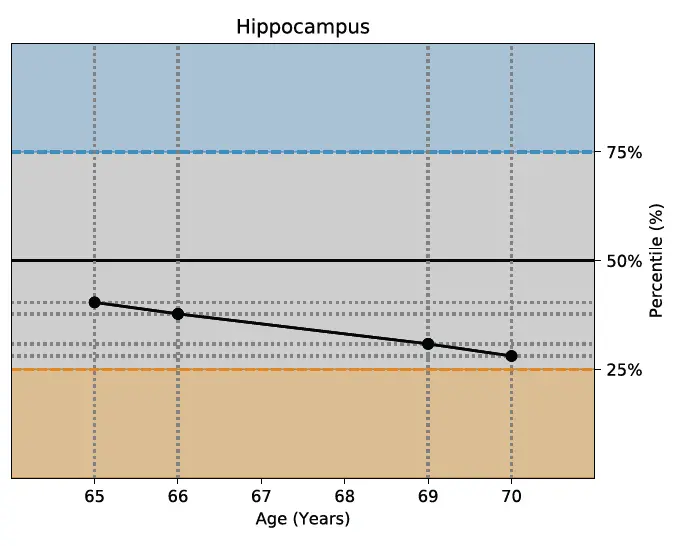
Longitudinal brain volumetry allows clinicians to compare measurements across multiple MRI examinations by tracking:
- Regional brain volume
- Whole-brain volume
- Percentile changes
- Structural progression
Objective longitudinal measurements may support ongoing patient monitoring as part of comprehensive clinical care.
When Should You Order Brain Volumetric MRI?
MRI and PET: Different Types of Information
MRI and PET imaging each contribute different information during the evaluation of neurodegenerative disease.
MRI primarily evaluates brain structure, while PET evaluates metabolic or molecular activity.
Depending on the patient's clinical presentation, these imaging modalities may be complementary rather than competing approaches.

Volumetric MRI vs PET Scans: Cost, Access, and Clinical Value
Real-World Clinical Examples
Published research and Neuroreader® case studies illustrate how objective brain volumetric analysis can complement the evaluation of patients with Alzheimer's disease and frontotemporal dementia.
Alzheimer's Disease Case Study
See how quantitative brain volumetry provided objective measurements that complemented routine MRI interpretation during the evaluation of a patient with Alzheimer's disease.
Frontotemporal Dementia Case Study
Explore a real-world example demonstrating how objective volumetric measurements provided supplementary volumetric measurements that were interpreted alongside the patient’s conventional MRI and broader clinical evaluation.
Integrating Quantitative MRI Into Clinical Practice
Neuroreader® works with routine high-resolution 3D T1-weighted MRI scans already acquired at many hospitals and imaging centers.
Flexible deployment options include:
PACS integration
Secure web-based access
Mediation server deployment
This may allow healthcare organizations to incorporate volumetric analysis into existing workflows when a compatible high-resolution 3D T1-weighted sequence is already included in the MRI protocol and meets applicable image-quality requirements.
How to Integrate Brain Volumetric Analysis into Existing PACS Workflows
Supporting Clinical Confidence With Objective Measurements
No imaging study should be interpreted in isolation.
Diagnosing neurodegenerative disease requires integrating patient history, neurological examination, cognitive testing, laboratory findings, imaging, and physician expertise.
Quantitative brain MRI adds another layer of information by providing standardized, objective measurements of brain structure.
When evaluating conditions such as Alzheimer's disease and frontotemporal dementia, these measurements can complement conventional MRI interpretation and support more informed clinical assessment.
Learn More About Neuroreader®
Neuroreader® is an FDA-cleared, CE-marked brain volumetric analysis software that automatically labels, visualizes, and quantifies 83 brain structures from compatible MRI studies, generating objective volumetric measurements in less than 10 minutes.
Whether your organization is evaluating patients with suspected neurodegenerative disease or looking to integrate quantitative brain volumetry into its imaging workflow, our team can help you explore implementation options, clinical applications, and onboarding resources.
Learn more about getting started with Neuroreader®