
Brain MRI Is a Powerful Tool, But Sometimes More Information Is Helpful
Magnetic resonance imaging (MRI) remains one of the most important tools for evaluating patients with neurological symptoms. Conventional MRI provides detailed anatomical images that allow radiologists and neurologists to assess brain structure, identify abnormalities, and support clinical diagnosis.
However, many neurological disorders develop gradually. Structural brain changes associated with neurodegenerative disease may be subtle, progressive, and difficult to quantify through visual assessment alone.
This is where brain volumetric analysis, an application of quantitative MRI post-processing, can provide additional insight.
What Is Quantitative MRI and Why Does It Matter?
Rather than relying solely on visual interpretation, quantitative brain volumetry objectively measures brain structures, compares them to age-adjusted normative data, and enables longitudinal comparison across multiple MRI studies.
While volumetric analysis is not appropriate for every patient, there are several clinical scenarios where it may provide valuable information to complement routine MRI interpretation and the broader clinical evaluation.
What Is Brain Volumetric MRI?
Brain volumetric MRI is an advanced image-processing technique that analyzes routine high-resolution 3D T1-weighted MRI scans to calculate the volume of specific brain structures.
Neuroreader® automatically segments and quantifies 83 brain regions in less than 10 minutes, producing standardized measurements that can complement radiologist interpretation.
For a deeper explanation of how quantitative brain analysis works, read our article on What Is Quantitative MRI (qMRI) and Why Does It Matter?

83
Brain volumes

<10 MIN
Automated analysis

3D T1
Compatible MRI Sequence
Neuroreader® does not independently diagnose, rule out, or differentiate neurological disorders. Results must be interpreted by a qualified healthcare professional together with conventional MRI findings and the complete clinical evaluation.
Clinical Scenarios Where Brain Volumetric MRI May Add Value

1. Patients Presenting With Memory Concerns
One of the most common reasons for ordering brain volumetric MRI is the evaluation of patients experiencing memory loss or cognitive decline.
Symptoms such as:
- Increasing forgetfulness
- Difficulty completing familiar tasks
- Problems with language
- Changes in executive function
- Confusion regarding time or place
may prompt clinicians to investigate potential structural brain changes.
Quantitative MRI provides objective measurements that can complement conventional MRI interpretation during the evaluation of these patients.
If you'd like to learn more about why identifying structural brain changes early is important, explore our article on How Early Detection of Neurodegenerative Diseases Improves Patient Outcomes.
How Early Detection of Neurodegenerative Diseases Helps Patient Outcomes

2. Suspected Mild Cognitive Impairment (MCI)
Mild Cognitive Impairment (MCI) represents an important stage between expected cognitive aging and dementia.
Not every patient with MCI develops Alzheimer's disease, making accurate assessment particularly important.
Because structural brain changes may be subtle during this stage, objective volumetric measurements may provide additional information alongside neurological examination, neuropsychological testing, and conventional imaging.
Longitudinal volumetric analysis may also assist clinicians when monitoring structural changes over time.

3. Evaluation of Suspected Alzheimer's Disease
Alzheimer's disease is characterized by progressive structural changes within the brain, particularly in regions associated with memory and cognition.
Brain volumetric MRI provides objective measurements of structures commonly evaluated during the assessment of neurodegenerative disease, helping clinicians quantify anatomical changes rather than relying solely on visual estimation.
When combined with the patient's history, cognitive testing, laboratory evaluation, and conventional MRI findings, quantitative brain volumetry can contribute additional objective information to the clinical assessment.
Clinicians evaluating Alzheimer's disease may also benefit from understanding how volumetric MRI compares with functional imaging techniques such as PET.
Volumetric MRI vs PET Scans: Cost, Access, and Clinical Value
4. Differentiating Neurodegenerative Disorders
Many neurodegenerative diseases share similar clinical symptoms.
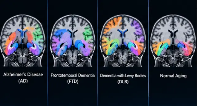
For example, patients with Alzheimer's disease and frontotemporal dementia (FTD) may both present with memory problems, behavioral changes, or cognitive decline.
Because different neurological conditions can affect different brain regions, objective volumetric measurements may provide additional structural information that complements the diagnostic process.
Quantitative MRI should always be interpreted within the context of the patient's complete clinical picture.
From Subjective to Objective: How Volumetric MRI Changes Brain Imaging

5. Longitudinal Monitoring
Neurological diseases often evolve gradually over months or years.
A single MRI provides valuable structural information, but comparing multiple MRI examinations may reveal meaningful changes that are difficult to appreciate through visual comparison alone.
Longitudinal brain volumetry allows clinicians to objectively compare measurements across serial MRI studies by tracking:
- Regional brain volume
- Whole-brain volume
- Percentile changes
- Structural progression over time
This information may support ongoing patient monitoring and clinical evaluation.
How Early Detection of Neurodegenerative Diseases Helps Patient Outcomes
6. Traumatic Brain Injury (TBI)
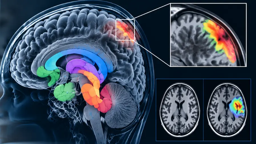
Patients with traumatic brain injury may experience persistent neurological symptoms despite relatively subtle findings on conventional imaging.
Quantitative MRI provides objective volumetric measurements and left-right asymmetry indices that may complement routine MRI interpretation during the evaluation of patients with a history of TBI.
Objective measurements may also be useful when comparing follow-up imaging over time.
Quantitative MRI in Traumatic Brain Injury: What Clinicians Should Know
7. Mesial Temporal Sclerosis (MTS)
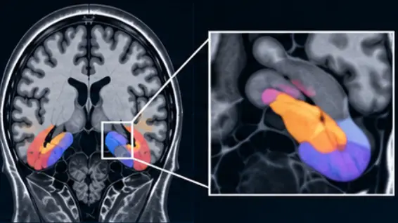
Mesial temporal sclerosis is one of the most common causes of medically refractory temporal lobe epilepsy.
Accurate identification of hippocampal atrophy is an important component of evaluating these patients.
Automated volumetric analysis provides standardized hippocampal measurements that can complement conventional MRI interpretation by objectively quantifying hippocampal volume and asymmetry.
Improving Detection of Mesial Temporal Sclerosis with Quantitative MRI
When Quantitative MRI May Not Be Necessary
Not every brain MRI requires volumetric analysis.
Routine examinations performed for:
- Acute trauma
- Intracranial hemorrhage
- Stroke evaluation
- Brain tumors
- Infection
may not require quantitative brain volumetry, depending on the clinical question.
Like any advanced imaging tool, quantitative MRI provides the greatest value when selected to answer specific clinical questions related to brain structure and neurological disease.
Integrating Quantitative MRI Into Clinical Practice
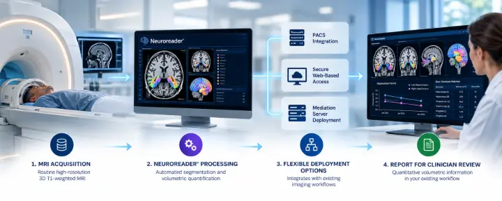
One advantage of Neuroreader® is that it works with routine high-resolution 3D T1-weighted MRI sequences already acquired at many hospitals and imaging centers.
Neuroreader® integrates with existing imaging workflows through flexible deployment options, including PACS integration, secure web-based access, and mediation server deployment.
When a compatible high-resolution 3D T1-weighted sequence is already available and meets image-quality requirements, an additional acquisition may not be necessary.
Learn more about implementation in our guide:
How to Integrate Brain Volumetric Analysis into Existing PACS Workflows
Objective Data to Complement Clinical Expertise
Brain volumetric MRI is not intended to replace physician judgment.
Instead, it provides standardized, objective measurements that can complement conventional MRI interpretation and support the broader clinical evaluation.
As neurological imaging continues to advance, quantitative brain volumetry is becoming an increasingly valuable component of precision medicine, providing clinicians with additional information that may help evaluate patients with suspected neurodegenerative disease, traumatic brain injury, mesial temporal sclerosis, and other neurological conditions.
To learn how objective measurements are changing modern neuroimaging, read:
From Subjective to Objective: How Volumetric MRI Changes Brain Imaging
Learn More About Neuroreader®
Neuroreader® is an FDA-cleared, CE-marked brain volumetric analysis software that transforms routine brain MRI into objective brain health insights by quantifying 83 brain structures in less than 10 minutes.
Healthcare organizations exploring quantitative brain imaging should also consider the operational and financial aspects of implementation.
Additional resources:
The Business Case for Offering Volumetric MRI in Imaging Centers
The Economics of Volumetric Imaging
How to Integrate Brain Volumetric Analysis into Existing PACS Workflows
If your organization is interested in incorporating quantitative brain volumetry into its imaging workflow, our team can help you explore implementation options, clinical applications, and onboarding resources.
Learn more about getting started with Neuroreader®